Gel electrophoresis
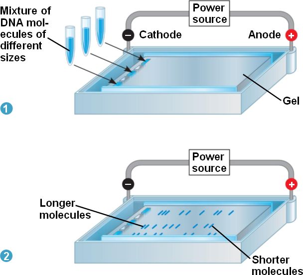
Gel electrophoresis takes advantage of DNA’s negative charge. DNA that has been subjected to restriction endonuclease digestion will be cleaved into fragments of different lengths. A solution containing different-size fragments to be separated is placed in a well.
A well is a depression at one end of the gel. The gel itself is usually a square or rectangular slab and consists of a buffer containing electrolytes and Agarose. The DNA solution containing fragments to be separated is mixed with a loading dye containing glycerol. The loading dye allows visualization of the DNA solution.Glycerol is a heavy molecule that causes the DNA solution to sink down into the well. The gel is loaded while it is submerged in a tray containing an electrolytic solution called the buffer.Using direct current, a negative charge is placed at one end of the gel where the wells are, and a positive charge is placed at the opposite end of the gel. The electrolyte solution conveys the current through the gel. The negatively charged DNA will migrate toward the positively charged electrode, with the shorter fragments migrating faster than the longer fragments, achieving separation. Small molecules found within the loading dye migrate ahead of all the DNA fragments. The shorter the fragment is, the faster it will travel because of its ability to navigate through the pores in the gel more easily than a large fragment can.